The School of Molecular & Cellular Biology is pleased to congratulate the 2026 winners of its “Life Inspiring” art competition, which celebrates the intersection of life sciences and art.
Students, staff, and faculty were encouraged to submit original works of art reflecting their scientific endeavors. Accepted mediums included graphic design, drawings, paintings, photography, and mixed media pictures. Winning artwork will be framed and displayed on the first floor of Burrill Hall.
Congratulations to this year's winners!
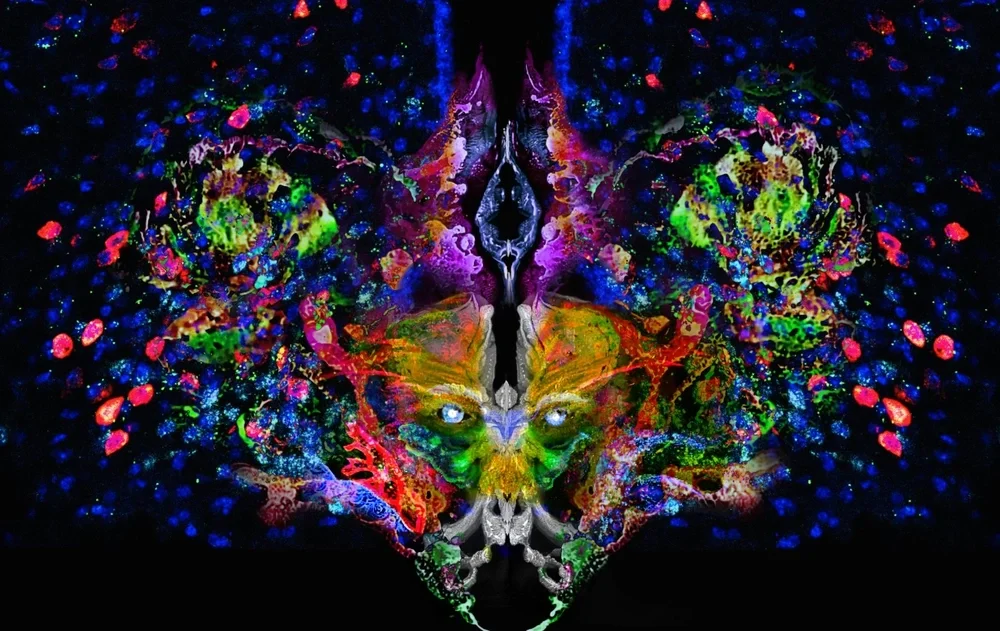
First Place: Face of Adaptation

Kerem Catalbas, Neuroscience PhD student, Sweeney Lab
This piece blends RNAscope confocal images from the mouse brain with watercolor textures I created by hand, then photographed and digitally layered into a single composition. The biological signals come from the arcuate nucleus, a small but powerful hub that helps the brain sense the body’s energy needs and adjust feeding and metabolism. Here, hunger-promoting AgRP neurons and satiety-promoting POMC neurons sit side by side, constantly negotiating balance. By transforming these cellular patterns into a symmetrical, mask-like face, I wanted adaptation to feel like a presence, something you can “meet,” not just measure. This piece reflects my research on how brain circuits flex in response to changing physiological demands to keep the body in balance.
Catalbas is also in the running for the The People's Artist competition presented by Art Form magazine and Johnny Depp.
Second Place: Forbidden Gummies: Clear, Colorful Critters and Cholera
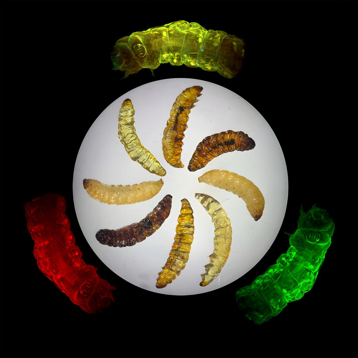
Jehdah Me'a'ofa Burton, member of the Grant Lab, microbiology PhD student
My research seeks to develop the waxworm Galleria mellonella as a model for studying the pathogenesis of Vibrio cholerae. All science aims to achieve clarity, but in my case, this is a literal endeavor—to render the whole waxworm transparent in order to visualize fluorescently-tagged bacteria. The central image displays waxworms as they might be found in the wild or in a bait shop, alongside representatives from various clearing attempts. These varying results remind me that scientific discovery is not only marked by error, but crucially molded by it. These “failed” amber waxworms, though unable to provide insight via fluorescence microscopy, still reveal nuances within the clearing procedure. On the periphery are fluorescence microscope images from transparent waxworms. Even without added tags, the tissues autofluoresce in striking colors. In spite of their unassuming nature, there is truly more to Galleria mellonella than meets the eye.
Third Place: Life in a Glass Jar
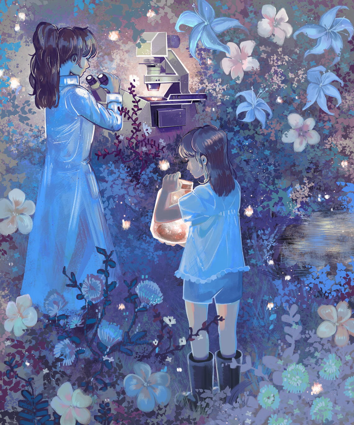
Abigail Finn, Microbiology PhD student, Whitaker Lab
I have a feeling that many people developed an interest in science for at least one similar reason, that the joy and wonderment in appreciating the natural world never escaped us since its ignition in our youth. Much of my research involves microscopic imaging, a technique I probably gravitated towards due to a preexisting fixation on visual art. Therefore, I wanted to evoke a painting by John Singer Sargent that I really enjoy called 'Carnation, Lilly, Lilly, Rose.' In my opinion, painters like Sargent deeply loved light since it opened the world to a painter's eyes in infinite variation. I too, have come to deeply appreciate light for the ways it contributes to our revelations of life's smallest fabrications, from bugs in a glass jar to bugs on a slide. This piece was painted by hand using an open-source digital painting program and a tabletop tablet and pen stylus.