OMX V2 Imaging platform includes super resolution technology providing spatial resolution far better than the diffraction limited performance in a normal epi-fluorescent microscope. The platform also includes the integrated capability for multiple cameras that can expose simultaneously for extremely fast, high signal-to-noise, live cell, and multiple wavelength imaging applications.
- Super-resolution Microscope
- Deconvolution Light Microscopy
- High Speed Imaging
- Power User Prices
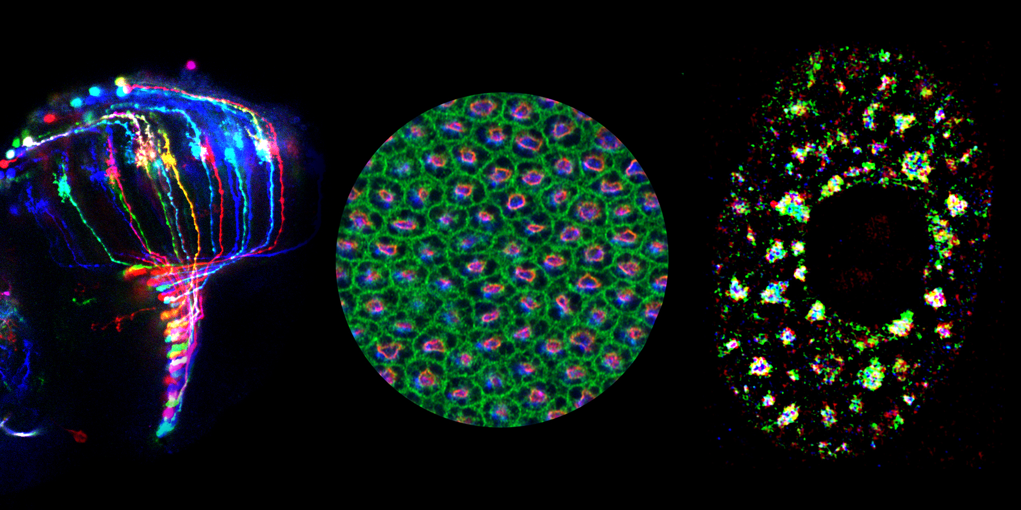
Super-resolution microscope ready for general users
We now are opening the new Applied Precision OMX structured illumination microscope for general use. This microscope offers roughly twice the resolution of conventional confocal or deconvolution microscopy, with 100 nm in x-y and ~200 nm z resolution.
A key feature of this technique is that it is essentially just an adaptation of conventional deconvolution microscopy so that you can use the same slides and dyes used in your normal light microscopy work.
We anticipate an hourly rate of ~$30/hr for use. However, training and an initial 20 hrs on the instrument will be free. Moreover, labs serious about use of this instrument can purchase a share of the service contract cost, which will reduce the effective hourly charge significantly.
The training path for this microscope will require initial training on the Personal Deltavision deconvolution microscope which is used to survey your slides and select regions of particular interest for examination with the OMX. Once users are proficient on the Deltavision they then will be trained on the OMX, which uses some of the same software packages as used on the Deltavision.
New deconvolution light microscopy system ready for general users
We are now opening the Applied Precision Personal Deltavision system for general use when OMX users are not using it. The Deltavision is arguably the best of the commercially available deconvolution systems. It is very easy to learn and typically offers better resolution and sensitivity than confocal systems for cell monolayers and other samples where light scattering is not a serious problem. The system is setup to acquire up to 4 fluorescent channels plus transmitted light. The four filter sets available are for fluorochromes with excitation/emission spectrum comparable to DAPI/Hoescht/Coumarin, GFP/FITC/Cy2/Al488, Rhodamine/Texas Red/Cy3/mRFP, Cy5.
We anticipate an hourly rate of $15/hr for use. Training will be free. Moreover, labs serious about use of this instrument can purchase a share of the OMX service contract cost, which will reduce the effective hourly charge significantly.
New high speed imaging for live cell microscopy
The OMX microscope has a second mode providing high speed, wide-field light microscopy 3D imaging at up to 10 3D reconstructions per second. We have a 2 camera system, so that we can collect GFP and mCherry simultaneously at these speeds. Even if you do not need rapid time sampling, the rapid data acquisition “freezes” cell movements during a given 3D data set. Moreover, because the two colors are collected simultaneously with each exposure there is zero spatial shift due to time delays between data acquisition of the two channels as experienced on other microscopes. Several labs using this system to study mitosis are “seeing” what they were not able to see before. This should be an excellent tool for correlating the localization of two proteins in live cells when these proteins are on moving structures.
We anticipate an hourly rate of ~$30/hr for use. However, training and an initial 20 hrs on the instrument will be free. Moreover, labs serious about use of this instrument can purchase a share of the service contract cost, which will reduce the effective hourly charge significantly.
The training path for this microscope again will be to first be trained to use the Personal Deltavision microscope which will be used to survey your slides and select regions of particular interest for examination with the OMX. Once users are proficient on the Deltavision, they will then be trained on the OMX, which uses some of the same software packages as used on the Deltavision.
Power user prices
To encourage and maximize usage on the facility microscopes I would like to introduce on an experimental bias an alternative mechanism for service charges.
The idea is that labs would be able to purchase a percentage of the total service contract or operating cost for the microscope and pay a flat fee for a fixed share of the available hours per year. By my calculations for heavy users this should drop the microscope cost per hour several fold.
My hope is that we can maximize instrument usage in a win-win situation for both the individual labs and the facility. Users will see greatly reduced costs while hopefully the facility will see increasing use with lowering of costs while recovering a larger fraction of service contract expenses. For instruments such as the new Deltavision and OMX, the cost of operating the microscopes is nearly independent of usage. Therefore it makes sense to try to reduce costs to maximize instrument usage as long as we can cover the service contracts. The idea is that if we can encourage greater hours on the instrument we can lower hourly charges dramatically. Even for the confocal, the only costs that scale with hours are the lasers that have a limited lifetime.
Therefore, the idea would be to offer time-shares. For the OMX, with a service contract of 30K per year, for $3000 per year a lab will have 10% of all hours available. Depending on demand we might have to adjust policies, but right now the offer would be that $3000 would buy a lab the equivalent of half a workday (M-F, 9-5, 4 hrs) per week and also 10% of all weekend and evening time (roughly another 6 hrs per week). This $3000 cost for a 10% share should be compared to a cost of 10 hrs/week x 50 weeks x $30/hr or $15,000 per year if equivalent microscope time was paid on an hourly basis.
Similarly for the confocal Zeiss 510, an estimated operating cost of 25K per year, $2500, would buy you 10% of microscope time (~10 hrs per week). This compares to a cost of $12,500 per year for 10 hrs per week x 50 weeks x $25/hr.
Please contact me (Andrew Belmont) if you are interested in this arrangement for either microscope.