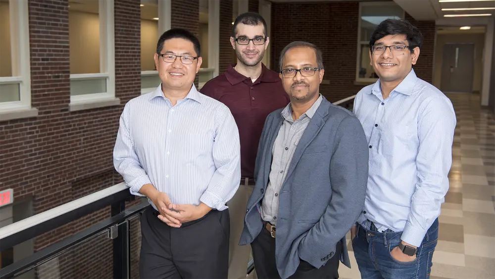
The Zhang and Pan labs recently published a joint paper in Nanoscale titled “Carbon dots with induced surface oxidation permits imaging at single-particle level for intracellular studies.”
Visualizing various components of a cell can be challenging and may require agents that offer multi-color emission. Some cellular components give off their own light and therefore any visualization technique requires a system that generates a bright signal. Additionally, in order to dissect cellular processes better, the technique needs to create different color signals.
“Carbon dots are a unique class of luminescent materials. Although they have been known for over a decade, they are recently attracting a lot of renewed attention because of their simple production chemistry, tunability and also they can be introduced into cells without causing any cellular damage,” said Dipanjan Pan, Associate Professor and Director of Professional Master’s Program in the Department of Bioengineering. Carbon dots have the added advantage that they can be made from an easily accessible range of materials using simple laboratory techniques.
The strong luminescent properties of carbon dots are based on the surface defects present at the nanoscale and also other surface characteristics. Several works from Pan Lab and others in past demonstrated that tweaking the surface components changes the color properties of these particles. “Although in this paper we only look at four colors, in reality we can create a diverse color range. This property will be useful in studying several cellular pathways,” according to Kai Zhang, Assistant Professor in the Department of Biochemistry.
The Zhang lab has always been interested in looking at particles that can be used for cellular imaging. “I have used quantum dots which are semiconductor nanocrystals to trace axonal transport of molecules in live neuronal cells. Kinetics of axonal transport, outlined by nanoparticles, have been used to study nerve disorders such as amyotrophic lateral sclerosis (ALS) and Alzheimer’s. However, nanoparticles need to undergo several modifications in order to be compatible with a biological system,” Zhang said.
Pan’s lab is interested in investigating next generation carbon dots with distinct multi-color emission for bio-imaging at cell, tissue and organ level. “We are looking to do research on materials that have immediate impact in biomedical fields. When I saw Kai’s work with quantum dots, I reached out and asked whether we could collaborate on the use of carbon dots for single particle imaging,” said Pan.
In the paper, the Pan lab used a catalytic process to change the surface of the carbon dots to render them more ‘oxidized’. “By using a catalyst that is ‘sacrificial’ in nature, we hit two birds with one stone. The catalyst by itself gets consumed during the synthesis but simultaneously oxidized the nanoscale surface,” said Pan. As the catalyst was being used up, it also caused changes that modified the groups on the surface of the carbon dots. Depending on the change, the dots were blue, green, yellow, or red. The lab then separated each category of dots and sent them to the Zhang lab for analysis.
“Our lab then immobilized these particles to determine their luminescent properties. Unlike most labs that look at them as a bulk, we focus on looking at the properties of individual particles. This allows us to accurately characterize them,” said Zhang.
According to Pan the biggest problem with carbon dots is their brightness. “The current techniques that we use result in a low yield of some of the biologically important fractions, e.g. red. Our future work will look at novel synthetic ways to increase the yield so that we can get a brighter signal free of biological interference.”
“The signal of the particles need to stand out against the background of the cell. This is a common challenge in the field, which is why carbon dots have not been investigated thoroughly. We believe that they have the potential to become powerful illumination materials that have applications in the fields of chemistry, biology, and material science,” Zhang said.
Both the labs agree that it has been a very productive collaboration. “We have complementary expertise and we currently have a huge pipeline of future projects. We believe that carbon dots will be the next generation of particles in imaging biological samples presenting excellent opportunities for clinical translation,” said Pan.
In addition to Pan and Zhang, bioengineering graduate student, Indrajit Srivastava; postdoctoral researchers Santosh K. Misra, Dinabandhu Sar, Aaron Schwartz-Duval; and biochemistry graduate research assistants, John Khamo and Vishnu Krishnamurthy; as well as Julio A. N. T. Soares of the Frederick Seitz Materials Research Laboratory, contributed to this work.
Photo by Steph Adams
