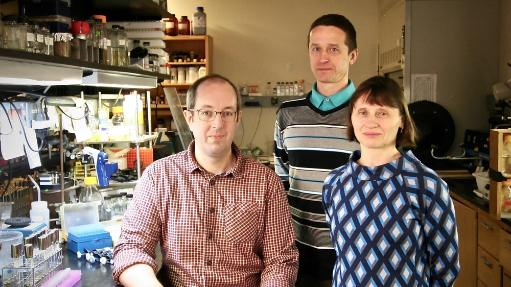
The latest paper by the Kuzminov lab describes the development of a highly sensitive method to probe the nature of DNA replication in E. coli. The findings were published in a paper titled “Near-continuously synthesized leading strands in Escherichia coli are broken by ribonucleotide excision” in the Proceedings in the National Academy of Sciences.
The information in biology textbooks often changes based on new experimental evidence. Biologists work to verify in vitro results, using purified enzymes in a test tube, with in vivo data, which look at cellular processes in live cells. Unfortunately, the unification of these two can sometimes take years due to unforeseen complications. Such an attempt has been made in the latest paper by the Kuzminov lab. The paper looks at DNA replication, a fundamental phenomenon that has been studied for over 50 years.
DNA consists of two strands wrapped around each other to form a double helix. The strands run in opposite directions; one strand runs from 5’ to 3’ while the opposite strand runs from 3’ to 5’. DNA replication takes place at the replication fork, which is formed by unwinding the DNA double helix. DNA polymerase, the enzyme responsible for synthesizing both daughter strands simultaneously, polymerizes nucleotides in a 5’ to 3’ direction. Therefore, the question arises: what do the daughter strands look like when they are first synthesized?
One daughter strand (“lagging”) must be made discontinuously because the replication machinery needs to stop and restart in order to maintain its overall 5’ to 3’ motion. However, the other “leading” strand could, in principle, be synthesized continuously. In vitro data support this hypothesis: when the DNA is synthesized in test tubes, the leading strand is recovered as a single unbroken strand.
On the other hand, in vivo experiments that involve extracting DNA replication intermediates indicate that the leading strand is synthesized discontinuously. Extracted from cells, the leading strand is comprised of short pieces — much like the lagging strand is. “Many believed that the in vivo results were artifacts arising from the cellular environment,” explained Dr. Andrei Kuzminov, Professor in the Department of Microbiology. “We remained skeptical of this interpretation and wanted to investigate the in vivo results thoroughly.”
Typically, DNA replication intermediates are analyzed using alkaline sucrose gradients, which dissociate the intermediates from the parent strands and separate them based on their size. Using this technique, the lab had previously shown that the leading strand was synthesized as low molecular weight fragments of approximately 1.2 Kb, suggesting discontinuous leading strand replication. These intermediates were later joined by the cell to form chromosomal-size DNA of 300 Kb.
However, Glen Cronan, a research scientist in the Kuzminov lab, has developed a high resolution alkaline sucrose gradient. “Using this technique we have observed intermediate molecular weight (5-25 Kb) fragments in the leading strand, which suggest "more" continuous replication than in the lagging strand, where the replication intermediates remained short, approximately 1 kb,” said Cronan.
The question then arises: why aren’t even higher molecular weight intermediates observed in vivo, indicating fully continuous replication? One of the problems in DNA replication is that the cell can accidentally incorporate ribonucleotides, which are the building blocks of RNA but not DNA, into the daughter DNA. “Ribonucleotides are incorporated 1 in 10,000 times. Additionally, since they outnumber the DNA nucleotides 200 to 1, ribonucleotide contamination is a serious problem,” said Elena Kouzminova, another research scientist in the lab. Therefore, the cell employs enzymes to nick the DNA and remove the ribonucleotides, a process that creates temporary breaks in the leading strand.
Additionally, Kouzminova has determined that the misincorporated ribonucleotides, which are sometimes not removed in vivo, react with the alkaline sucrose gradients thereby splitting the longer intermediates. "We realized that to detect the full length replication intermediates in the leading strand, we needed to change the method,” said Kuzminov. “Dr. Cronan has developed neutral formamide-urea sucrose gradients and used them to demonstrate the nearly-continuous replication on the leading strand in vivo.”
“This paper explains why there seemed to be such a massive contradiction between in vivo and in vitro results,” said Cronan. “Ribonucleotide contamination would not have occurred in in vitro experiments, which generated very clean results. On the other hand, newly synthesized DNA in vivo is quite "dirty" and needs to be cleaned before it becomes chromosomal DNA."
"This is a rare story with a happy ending," concluded Kuzminov. "It is satisfying to find that both in vitro and in vivo studies were right. The critical difference was this recently-discovered DNA contamination with RNA nucleotides that nobody knew about back in the sixties and seventies when the bulk of research on DNA replication intermediates was performed.”
