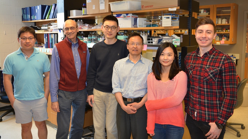
The latest paper by the Shapiro lab looks at the effect of mutations in the estrogen receptor on the growth and spread of breast cancer cells. The findings were published in a paper titled "Estrogen-independent Myc overexpression confers endocrine therapy resistance on breast cancer cells expressing ERαY537S and ERαD538G mutations" in Cancer Letters.
Metastatic cancer occurs when cancer cells cut themselves loose from the primary site, circulate in the blood stream, and anchor themselves in a secondary location. This process is responsible for the lethality associated with cancer. In the case of breast cancer, growth and metastasis is stimulated by binding of the hormone estrogen to its receptor. Treatment usually involves preventing cells from making estrogen or preventing the binding of estrogen to its receptor. However, cells often become resistant to these drugs.
“It has become apparent that in about one-third of patients with metastatic breast cancer the estrogen receptor is mutated and remains on even in the absence of estrogen,” said Dr. David Shapiro, Professor in the Department of Biochemistry. The two point mutations most commonly associated with estrogen receptor in breast cancer are ERαY537S and ERαD538G. “Sadly, patients whose metastatic tumors contain these mutations die more rapidly. Therefore, we wanted to understand why tumors with these mutations are so aggressive.”
The lab found that the mutations affect the cancer-causing protein, or oncogene, Myc, and enhance the metastatic ability of the cancer cells. Myc has been been extensively studied for its role in cell proliferation and has been shown to be important to the growth of many types of cancer. “Normal estrogen receptors require estrogen in order for them to bind to the enhancer region of Myc, increasing its production. However, the mutated receptors bind to the Myc enhancer even in the absence of estrogen, leading to continuous Myc production,” explained Liqun Yu, a graduate student in the Shapiro lab.
The lab also showed that the mutations increase the metastatic ability of the cancer cells. However, they discovered that this phenomenon is independent of Myc. Historically, metastasis has been difficult to model in the laboratory. As a result, researchers primarily focus on the ability of the cancer cells to dissociate from the primary site. However, Dr. Chengjian Mao in the Shapiro lab developed a novel assay to quantify the number of cells that can colonize a secondary location after breaking away from the primary tumor.
The assay looks at three steps involved in metastasis: invasion of the cancer cells through a protein-coated membrane, dissociation from the primary site, and rebinding to the secondary site. “We load the cells on a protein-coated membrane in a well. Cancer cells have the ability to digest this protein and go through to the underside of the membrane. They detach from the underside of the membrane and rebind to the bottom of the well. We can then quantify how many cells rebind to the bottom of the well,” said Yu. “The results are exciting because the ability of the cancer cells to cut through the membrane, detach, and go to the second site seems to be closely related to their ability to spread in a mouse,” added Shapiro.
Using this assay the lab is currently looking at what other proteins are important to the metastatic process. “Because it has been extremely difficult to assay, very few current cancer drugs target metastasis. Now we have the ability to identify small molecules that can affect not just the growth of cancer cells but also their ability to spread to other sites,” said Shapiro.
