HIV-1 affects and kills millions of people globally, but not enough information exists regarding the types of cells that HIV-1 targets in different tissues or the virus’s mechanism of spreading throughout the body. Viruses often ride the body’s circulatory systems to scatter throughout an organism and appear in different tissues to infect cells; however, not much is known about HIV-1’s mechanism of infection at the cellular level within those tissues. The Kieffer lab aims to provide insight into this topic by having recently published a study called “Mechanisms of virus dissemination in bone marrow of HIV-1-infected humanized BLT mice” in the journal eLife.
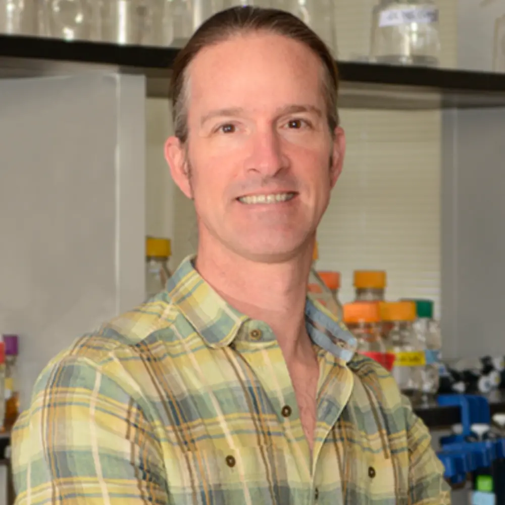
Collin Kieffer, Assistant Professor in the Department of Microbiology, is interested in bone marrow cell response to HIV-1 because he believes that even though bone marrow cells are underappreciated in HIV-1 studies, they could be extremely important for virus dissemination. He explained, “The greatest amount of virus production occurs when immune cells activate. Bone marrow is not a highly activated tissue, so we tend not to see as many viruses there as we would in, for example, gut-associated lymphoid tissue. But even though the density of infection in the body is not nearly as high in bone marrow as in it is other tissues, its role in spreading the virus is important. As a lymph station (in a system that regularly circulates cells throughout the body), it’s an attractive route for the virus to take.”
The lab conducted the study on mice that were genetically modified to express human-like immune systems. This animal model provided conditions that helped the researchers closely observe how virus activity in human patients might progress. Getting access to human HIV-1-infected tissues is usually costly and difficult, so this model satisfactorily served the lab’s purpose instead.
For imaging bone marrow cells, the lab combined several techniques—tissue clearing, immunofluorescence (IF), and light sheet fluorescence microscopy (LSFM). This combination allowed the researchers to view a large portion of tissue at once, and the cellular-level resolution—specific enough to observe a fixed volume of individual cells—showed exactly how multiple HIV-1-infected cells were distributed throughout a population of cells, as well as which types of cells were frequently targeted by HIV-1. Figures obtained from this imaging can help researchers delineate the steps of HIV-1 infection by comparing the chronology of multiple in-process infections.
Kieffer’s lab also used electron tomography (or ET, a 3-dimensional form of electron microscopy) to provide resolution of lymphoid tissues at the subcellular level, which can clearly show virus interaction with cellular membranes. However, the higher resolution sacrifices the ability to monitor viral activity among a large population of cells, meaning that only a few infected cells can be monitored at once.
The discrepancies in the cellular target scales of imaging between IF with LFSM (large-scale imaging) and ET (small-scale imaging) is why the lab used both techniques to describe their experimental observations (calling the process “multi-scale imaging”). In going from large-scale to small-scale imaging, the lab can identify at which cells infection occurs in a population and then zoom into those infected cells to understand how the infection process occurs. In Kieffer’s words, “Once we know who to look for and where to look for them, then we can go and look for the needles in the haystack more intelligently and understand the structural mechanisms that occur during infection. That means that going from big to small will help us understand HIV dissemination in tissues.”
This multi-scale imaging approach surprisingly showed that a mixture of two types of infection processes occurred: (1) direct cell-to-cell transmission, where one infected cell contacted another uninfected cell and deliberately transferred the virus, and (2) the random process of free viruses diffusing away to find new targets. Kieffer said, “This finding is controversial in the field because, in cultured cells, viral transmission from cell to cell is much more efficient than free viruses—that’s also the assumption of how it occurs in the body. But we found that equivalent levels of each process occurred in tissues.”
In addition to observing a mixture of those two infection mechanisms in tissues, the lab also surprisingly found that macrophages in bone marrow predominantly produced viruses. In the rest of the body, HIV-1 predominantly targets T-cells (specifically CD4+ T-cells) for infection and dissemination, and the lab expected those same cells to be targeted in bone marrow as well. However, macrophages comprised the majority of virus-associated cells in bone marrow. The reason the lab even focused on macrophages at all is because “it’s becoming increasingly clear that macrophages are important for HIV infection and viral dissemination, even though there’s not much evidence for it in tissues,” Kieffer explained. “So we wanted to see how involved macrophages are in virus dissemination in bone tissues for this study.”
“Bone marrow is the main site of hematopoiesis in the body (or the main spot where immune development occurs),” Kieffer said. Therefore, since macrophages are immune cells that originate in bone marrow and are tasked with circulating to other parts of the body, then virus production in macrophages serves as a dangerously effective method of spreading the virus to different tissues. Additionally, once a macrophage produces a virus, it may not release it immediately. It could rather store the virus inside itself before circulating to an area of greater cell population density and releasing it at that location to potentially infect more cells.
These findings encourage the lab’s search for answers about HIV-1 infection mechanisms in bone marrow cells and virus dissemination to other tissues. “This paper is part of our lab’s larger goal to visualize interactions between the host immune system and pathogens at all levels of volume resolution before we go, bit-by-bit, into the finer details,” said Kieffer.
Future work in this area will consist of moving on from studying mouse cells in genetically-modified animal models to imaging human patient cells directly. Kieffer said, “These methods are translational into human patients and any type of tissue. We also want to be the first to look at human patient samples and see what’s happening in large volume within those tissues as well.”
This paper was published by the Kieffer lab in collaboration with Dr. Dong Sung An from the UCLA Department of Nursing and with post-doctoral advisor Pamela Bjorkman from Caltech. Press release provided by Laura Tanase.
