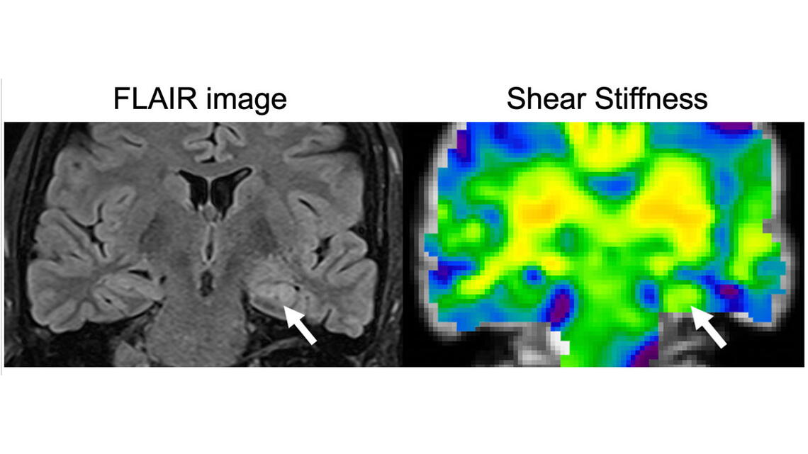
A new study uses magnetic resonance elastography to compare the stiffness of the hippocampus in patients who have epilepsy with healthy individuals. The technique can improve the detection and characterization of the disease.
The study “Hippocampal stiffness in mesial temporal lobe epilepsy measured with MR elastography: Preliminary comparison with healthy participants” was published in NeuroImage: Clinical. The work was done through a collaboration with the Carle Neuroscience Institute and the Beckman Institute for Advanced Science and Technology at the University of Illinois Urbana-Champaign.
Mesial temporal lobe epilepsy is the most common form of epilepsy that is resistant to medication. Unfortunately, current detection methods, which include magnetic resonance imaging, can only visualize the epilepsy-induced changes in the brain after significant damage has occurred.

“The structural changes in the brain, in response to seizures, causes the death of neurons and the formation of scar tissue,” said Graham Huesmann, a neurologist at Carle and a research assistant professor of molecular and integrative physiology, who is a part-time faculty member at the Beckman Institute. “By the time we see any changes on the MRI, the disease is pretty advanced. We wanted to detect these changes earlier using MRE.”
Early detection of these changes is critical for the disease, especially because it causes very mild symptoms in the beginning stages. “It starts with a feeling of déjà vu, which becomes more common as the disease progresses. Eventually it develops into a form that is medication resistant,” Huesmann said. “MRE allows us to detect these changes earlier affording us the opportunity to change the course of treatment.”
The researchers are now focusing on how to optimize the technique and also look at other types of epilepsy. “All of our imaging techniques currently depend on looking at brain chemistry and the static images of the brain,” Huesmann said. “Using MRE to see how the brain jiggles is an exciting way to approach this problem. It is also an inexpensive technique and can therefore be used by anyone.”
