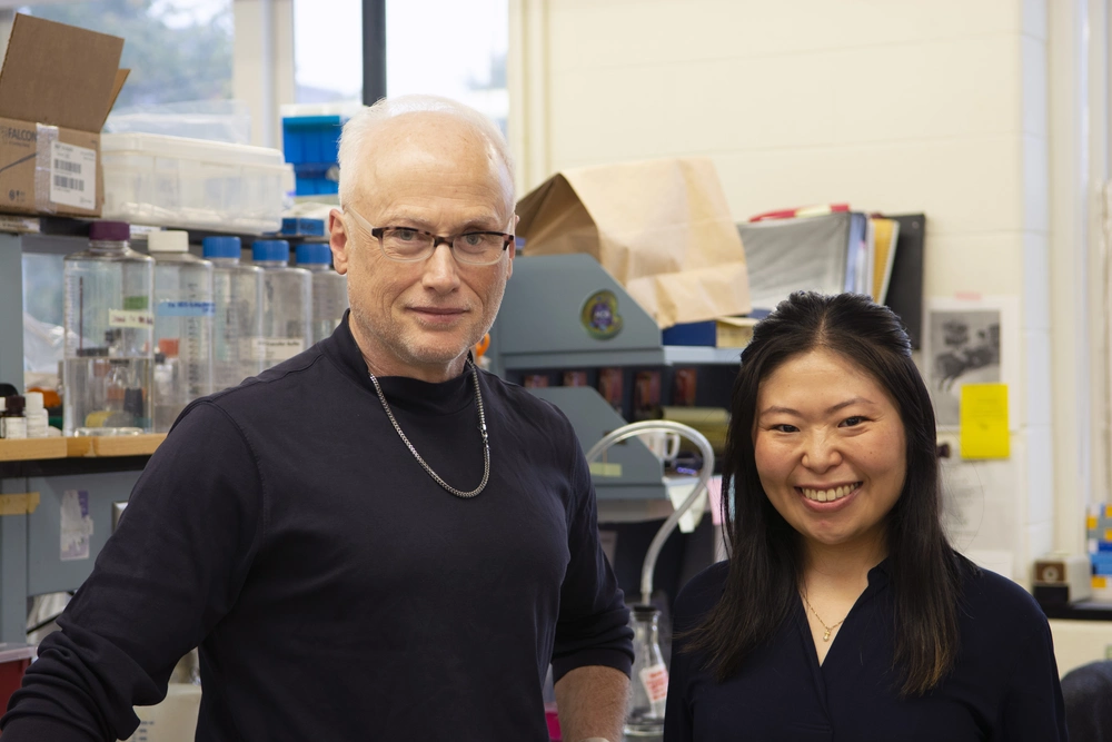
University of Illinois researchers have elucidated a mechanism whereby gastric cells detect and respond to toxin infiltration within the cell’s powerhouse, the mitochondria.
The Blanke lab, within the Department of Microbiology in the School of Molecular & Cellular Biology, studies the gastric pathogen Helicobacter pylori, or H. pylori, which chronically infects the human stomach as the single most important risk factor for development of stomach cancer. Exactly how the bacterium can sustain long-term infection in the hostile environment of the human stomach is poorly understood, researchers said.
To promote long-term infection, H. pylori generate a portfolio of virulence factors, including a protein toxin called Vacuolating cytotoxin A, or VacA. Upon release from the pathogenic bacterium, VacA has the capacity to enter host gastric cells, resulting in fundamental changes to the stomach, which promote long-term infection as a predominant risk factor for malignancy, according to researchers.
“We have learned that H. pylori secrete VacA to reprogram metabolism in the stomach, which has a number of functions that require energy. This toxin targets the mitochondria, the powerhouse of the cell, and the net result is that the loss of energy seems to cripple the cell’s ability to respond effectively to the presence of the pathogen,” said Dr. Steven Blanke, professor of microbiology and principal investigator of the study.
Earlier work suggested that one of the primary roles of VacA during infection was to directly damage gastric tissue. However, recent work surprisingly revealed that direct infusion of purified VacA continuously into stomachs of mice for an extended period did not result in destruction of the gastric tissue as previously believed, said Ami Seeger, PhD candidate in microbiology and first author of the paper. This prompted scientists to theorize that the gastric cells possess a mechanism that limits and protects cells from damage resulting from the targeting of mitochondria by VacA. The authors sought to uncover the mystery mechanism.
Their findings were described in the paper, “Host cell sensing and restoration of mitochondrial function and metabolism within Helicobacter pylori VacA intoxicated cells,” published recently in mBio, the flagship journal of American Society for Microbiology.
Deconstruction of VacA-damaged mitochondria in gastric cells as an unexpected mechanism to ultimately restore energy metabolism
The researchers discovered that human stomach cells can detect VacA and reduce mitochondrial damage caused by the toxin.
“We realize there is a way to sense the abnormal energy status within cells and to respond accordingly which involves, surprisingly, deconstruction of mitochondrial structure,” Seeger said.
During VacA infiltration, cellular energy is depleted, which signals the host energy sensor protein called AMPK. Seeger and colleagues found that AMPK is an exquisitely responsive sensor that recognizes the low energy status within H. pylori infected cells. Activation of AMPK sounds the alarm for cellular recruitment of repair machinery that works to restore mitochondrial structure and function while simultaneously removing mitochondrial-associated toxin.
Deconstruction of mitochondrial structure requires a protein factor Drp-1, that functions as molecular scissors to cut out VacA-damaged regions of mitochondria.
When Drp-1 is recruited to the damaged area of the mitochondria, it orchestrates the deconstruction of the organelle to isolate compromised areas from the overall mitochondrial network, resulting in the extraction of VacA from mitochondria.
“Temporarily shutting off the power within cells seems to be required for repair of the damaged mitochondrial cable network,” said Blanke. Ultimately, a restored network results in the reestablishment of the energy grid necessary for sustaining gastric cell viability.
Remaining mysteries and future directions
Deconstruction of the mitochondrial network was somewhat of a surprise to the investigators because mitochondrial fragmentation within cells affected by VacA has generally been considered to be a lethal consequence to cellular stress.
“We now realize that mitochondrial deconstruction is a cellular response required to isolate and subsequently degrade damaged mitochondrial regions,” Blanke said.
The results provide new insights into how mammalian cells deal with the problem of detecting and clearing foreign protein toxins that have gained access to the intracellular environment of host cells and are not susceptible to antibody-mediated immune responses.
“Instead of detecting the protein toxin itself, our data suggest that cells sense the damaging action of the toxin during infection. The process of sensing and responding to pathogenic proteins is called the ‘guard hypothesis,’ which has been predominantly explored in the plant immunity field. In contrast, our understanding of these types of mechanisms in mammalian cells is in the early stages,” Seeger said.
These studies revealed the unexpected findings that VacA-mediated mitochondrial fragmentation has a protective effect on mitochondrial health. However, the fate of VacA following extraction from mitochondria remains to be identified. Ongoing work in the Blanke lab hints that following extraction from mitochondria, VacA is trafficked to degradative cellular compartments, which, as yet, have not been identified. The authors are curious as to the nature of whether similar mechanisms exist to neutralize other intracellular protein toxins within host cells.
This work was supported by the National Institutes of Health and Alice Helm Fellowship.
